

The Products Of Meiosis Are:
Learning Objectives
- Draw the chromosomal makeup of a jail cell using the terms chromosome, sister chromatid, homologous chromosome, diploid, haploid, and tetrad
- Recognize the part and products of mitosis and meiosis
- Compare and contrast the behaviors of chromosomes in mitosis and meiosis
- Recognize when cells are diploid vs. haploid
- Predict DNA content of cells in different phases of mitosis and meiosis
- Recollect and depict the phases of the cell cycle
- Chronicle the prison cell cycle stages to changes in Deoxyribonucleic acid content
The Jail cell Segmentation Wheel

Cell division cycle, effigy from Wikipedia. Cells that stop dividing exit the G1 phase of the cell cycle into a then-called G0 state.
Cells reproduce genetically identical copies of themselves by cycles of cell growth and division. The cell bicycle diagram on the left shows that a cell division cycle consists of 4 stages:
- G1 is the period after cell partitioning, and before the kickoff of Dna replication. Cells abound and monitor their environment to decide whether they should initiate some other round of jail cell sectionalization.
- South is the period of Deoxyribonucleic acid synthesis, where cells replicate their chromosomes.
- G2 is the period betwixt the end of DNA replication and the outset of cell partitioning. Cells cheque to make sure Dna replication has successfully completed, and make any necessary repairs.
- One thousand is the bodily menstruation of cell segmentation, consisting of prophase, metaphase, anaphase, telophase, and cytokinesis.
Chromosomes
Chromosomes were kickoff named by cytologists viewing dividing cells through a microscope. The modern definition of a chromosome now includes the office of heredity and the chemical composition. A chromosome is a Deoxyribonucleic acid molecule that carries all or part of the hereditary information of an organism. In eukaryotic cells, the DNA is packaged with proteins in the nucleus, and varies in construction and advent at dissimilar parts of the cell bike.
Chromosomes condense and become visible by light microscopy as eukaryotic cells enter mitosis or meiosis. During interphase (G1 + S + G2), chromosomes are fully or partially decondensed, in the form of chromatin, which consists of Deoxyribonucleic acid wound around histone proteins (nucleosomes).

In G1, each chromosome is a single chromatid. In G2, later DNA replication in S phase, as cell enter mitotic prophase, each chromosome consists of a pair of identical sis chromatids, where each chromatid contains a linear DNA molecule that is identical to the joined sister. The sis chromatids are joined at their centromeres, as shown in the image below. A pair of sis chromatids is a single replicated chromosome, a single package of hereditary data.
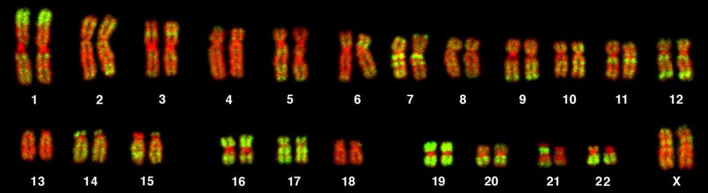
Human being karyotype "painted" using fluorescent Dna probes. These mitotic chromosomes each consist of a pair of sister chromatids joined at their centromeres. The images of the homologous chromosome pairs (e.thou., 2 copies of chromosome one) have been lined up next to each other. Image from Bolzer et al., (2005) 3-Dimensional Maps of All Chromosomes in Human Male Fibroblast Nuclei and Prometaphase Rosettes. PLoS Biol three(v): e157 DOI: 10.1371/journal.pbio.0030157
Ploidy
Humans are diploid, meaning we take ii copies of each chromosome. We inherited one copy of each chromosome from other mother, and one copy of each from our father. Gametes (sperm cells or egg cells) are haploid, pregnant that they have simply one consummate set of chromosomes.
Chromosomes that do not differ between males and females are called autosomes, and the chromosomes that differ between males and females are the sex chromosomes, Ten and Y for virtually mammals. Humans most commonly have 22 pairs of autosomes and i pair of sex chromosomes (Twenty or XY), for a total of 46 chromosomes. We say that humans have 2N = 46 chromosomes, where N = 23, or the haploid number of chromosomes.
Cells with complete sets of chromosomes are called euploid; cells with missing or extra chromosomes are called aneuploid. The virtually common aneuploid status in people is variation in the number of sexual practice chromosomes: XO (having but i re-create of the 10), Xxx, or XYY. Having no X chromosome results in early embryonic death.
The two copies of a particular chromosome, such as chromosome 1, are called homologous.The karyotype image above shows the homologous pairs for all the autosomes. Homologous chromosomes are not identical to each other, unlike sister chromatids. They oftentimes take dissimilar variants of the same hereditary information – such every bit blue eye color vs brown eye color, or blood type A versus claret blazon B.
Mitosis
Mitosis produces 2 girl cells that are genetically identical to each other, and to the parental jail cell. A diploid jail cell starts with 2N chromosomes and 2X DNA content. Afterward Deoxyribonucleic acid replication, the cells is still genetically diploid (2N chromosome number), only has 4X Dna content because each chromosome has replicated its DNA. Each chromosome at present consists of a joined pair of identical sister chromatids. During mitosis the sis chromatids split and get to opposite ends of the dividing jail cell. Mitosis ends with two identical cells, each with 2N chromosomes and 2X DNA content. All eukaryotic cells replicate via mitosis, exceptgermline cells that undergo meiosis (run across below) to produce gametes (eggs and sperm).
- prophase – chromosomes condense; each chromosome consists of a pair of identical sister chromatids joined at the centromere.
- metaphase – chromosomes line upwards at the eye of the prison cell, along the plane of jail cell partition, pushed and pulled by microtubules of the spindle apparatus
- anaphase – sis chromatids split up and migrate towards opposite ends of the cell
- telophase – chromatids cluster at contrary ends of the cell and brainstorm to decondense
- cytokinesis – the membrane pinches in to divide the two daughter cells
Here is a simplified diagram illustrating the overall process and products of mitosis:

Source: Wikimedia Commons (https://commons.wikimedia.org/wiki/File:MajorEventsInMeiosis_variant_int.svg)
Questions or points to ponder or notation about the figure above (answers at bottom of page):
- are the two girl cells the same or different from each other, and from the parent prison cell at the start?
- why does the cartoon analogy of the chromosomes change (from a single rod to joined double rods) afterward Dna replication, and again (back to single rods) during mitosis?
- does the figure show 2 different chromosomes or a single pair of homologous chromosomes?
- can haploid cells undergo mitosis? what about triploid cells (cells that take 3N chromosomes)?
This animation below shows the packaging of DNA and condensation of chromosomes as a cell undergoes mitosis.
The video narration has a major error at time 1:22: chromosomes exist throughout the entire cell cycle (at all times in a cell'south life); they are visible in their condensed form only during mitosis and meiosis.
Meiosis
This is a special sequence of two jail cell divisions that produces haploid gametes from diploid germline cells. It starts with a diploid jail cell that has undergone chromosomal Deoxyribonucleic acid replication: 2N chromosomes, 4X DNA content. Two successive divisions, with no additional Deoxyribonucleic acid replication, results in 4 haploid gametes: 1N chromosomes, 1X Deoxyribonucleic acid content.
NOVA has a good interactive side-by-side comparison of mitosis and meiosis on this folio: How cells divide
Meiosis sets the stage for Mendelian genetics. Students need to know that most of the genetics action occurs in the get-go meiotic division:
- homologous chromosomes pair upwardly and align cease-to-cease (synapsis) in prophase I
- crossing over occurs between homologous chromosomes in prophase I, before chromosomes line up at the metaphase plate
- homologous chromosomes separate to daughter cells (sister chromatids exercise not separate) in the first division, creating haploid (1N) cells
- the separation of each pair of homologous chromosomes occurs independently, and so all possible combinations of maternal and paternal chromosomes are possible in the ii daughter cells – this is the ground of Mendel's Law of Independent Assortment
- the start partitioning is when daughter cells become functionally or genetically haploid
The last indicate appears to be the most difficult for students to grasp. Consider the X and Y chromosomes. They pair in prophase I, and so separate in the outset division. The daughter cells of the first meiotic division have either an 10 or a Y; they don't have both. Each cell now has but 1 sex chromosome, like a haploid cell.
One mode of thinking about ploidy is the number of possible alleles for each cistron a cell can have. Correct after meiosis I, the homologous chromosomes have separated into different cells. Each homolog carries 1 re-create of the gene, and each gene could be a different allele, only these two homologs are now in 2 different cells. Though information technology looks like there are two of each chromosome in each cell, these are duplicated chromosomes; ie, it is 1 chromosome which has been copied, then there is but i possible allele in the cell (just two copies of it).
The 2nd meiotic sectionalisation is where sister (duplicated) chromatids separate. It resembles mitosis of a haploid jail cell. At the start of the 2nd segmentation, each jail cell contains 1N chromosomes, each consisting of a pair of sister chromatids joined at the centromere.
Hither is a simplified diagram illustrating the overall process and products of meiosis:
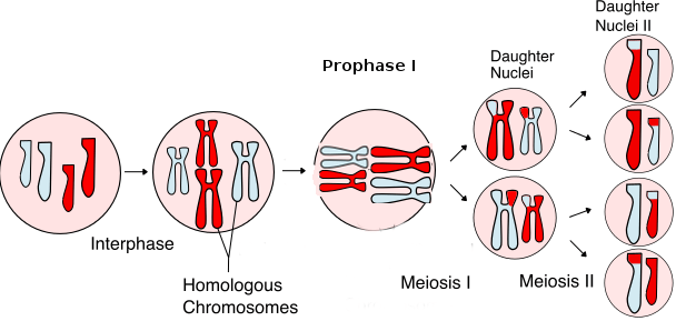
Meiosis Overview from Wikipedia by Rdbickel
And hither is a video that walks through the steps of meiosis:
It is very of import that yous recognize how and why cells become haploid after meiosis I.
To confirm for yourself that you understand meiosis, work through i or more of these interactive tutorials:
- The U. Arizona Cell Biology Project's Meiosis tutorial has a click-through blitheness of meiosis, with x idea-provoking problem questions.
- Jung Choi's interactive wink tutorial, programmed past Pearson, uses human chromosome 7, with wild blazon and cystic fibrosis alleles for CFTR, to track segregation through meiosis, with and without crossing over: Meiotic Segregation tutorial
Chromosomes, chromatids, what is the difference and how many chromosomes are there at different times of the prison cell cycle and after mitosis and meiosis?
Chromosomes by definition contain the Deoxyribonucleic acid that makes upwards the fundamental genome of the jail cell. In a prokaryote, the genome is usually packaged into i round chromosome consisting of a circular Deoxyribonucleic acid molecule of a few 1000000 base of operations pairs (Mbp). In eukaryotes, the genome is packaged into multiple linear chromosomes, each consisting of a linear Dna molecule of tens or hundreds of Mbp. Chromosomes exist at all unlike phases of the cell wheel. They condense and become visible to light microscopy in prophase of mitosis or meiosis, and they decondense during interphase, in the class of chromatin (DNA wrapped effectually nucleosomes, similar "beads on a string").
The chromosome number, N, in eukaryotes, refers to the number of chromosomes in a haploid cell, or gamete (sperm or egg prison cell). Diploid cells (all the cells in our torso except our gametes) have 2N chromosomes, because a diploid organism is created past spousal relationship of 2 gametes each containing 1N chromosomes. In terms of chromosome number (ploidy), information technology's useful to think of chromosomes equally packages of genetic data. A pair of sis chromatids is i chromosome because information technology has genetic information (alleles) inherited from only one parent. A pair of homologous chromosomes, each consisting of a single chromatid in a daughter cell at the stop of mitosis, has alleles from the begetter and from the mother, and counts equally two chromosomes.
This chromosome number stays the aforementioned later chromosome replication during Southward phase: each chromosome entering cell partition now consists of a pair of sis chromatids joined together at the centromere. Then in mitosis, the sis chromatids of each chromosome separate, and so each daughter cell receives one chromatid from each chromosome. The effect of mitosis is two identical girl cells, genetically identical to the original jail cell, all having 2N chromosomes. So during a mitotic cell bicycle, the DNA content per chromosome doubles during S stage (each chromosome starts as one chromatid, then becomes a pair of identical sister chromatids during S stage), merely the chromosome number stays the same.
A chromatid, then, is a single chromosomal Deoxyribonucleic acid molecule. The number of chromatids changes from 2X in G1 to 4X in G2 and back to 2X, simply the number of chromosomes stays the aforementioned.
The chromosome number is reduced from 2N to 1N in the first meiotic segmentation, and stays at 1N in the second meiotic sectionalisation. Because homologous chromosomes carve up in the kickoff division, the girl cells no longer take copies of each chromosome from both parents, then they have haploid genetic information, and a 1N chromosome number. The second meiotic division, where sister chromatids separate, is like mitosis. Chromosome number stays the same when sister chromatids split up.
Using the data above, compare these two simplified diagrams of mitosis and meiosis to visualize why cells are haploid after meiosis I. Specifically, compare the chromosomes in cells at the end of mitosis vs the end of meiosis I, recognizing that the diagram of mitosis tracks only a single pair of homologous chromosomes, whereas the diagram of meiosis tracks two pairs of homologous chromosomes (one long chromosome and short chromosome):

Meiosis Overview from Wikipedia by Rdbickel
The video below is geared toward a loftier schoolhouse audience, simply it does present a helpful way for recognizing how many chromosomes are present in a cell (and thus the ploidy level of that prison cell). While watching, meet if you can recognize why the products of meiosis 1 are haploid cells:
Answers to questions most the mitosis figure:
- The ii daughter cells are the same as each other, and same as the parental cell
- Each rod represents a chromatid, and Deoxyribonucleic acid replication results in two sister chromatids joined at their centromeres. Mitosis separates the sister chromatids.
- A single pair of homologous chromosomes. Red and blueish are chromosomes inherited from the male and female parents.
- Whatsoever cell tin can divide by mitosis – haploid, triploid, even aneuploid cells.

The Products Of Meiosis Are:,
Source: https://bioprinciples.biosci.gatech.edu/module-4-genes-and-genomes/4-1-cell-division-mitosis-and-meiosis/
Posted by: petersonhadioncoulne1959.blogspot.com
0 Response to "The Products Of Meiosis Are:"
Post a Comment